

Lung Cancer Classification using a Custom Residual CNN (PyTorch)
Project Overview
This project presents a deep learning framework for multi-class lung cancer classification from histopathological images using a custom Residual Convolutional Neural Network (ResCNN) implemented entirely in PyTorch.
The model distinguishes between healthy lung tissue and two major lung cancer subtypes while leveraging ResNet-inspired residual learning to improve optimization stability, gradient propagation, and convergence speed. The objective is to establish a lightweight yet effective baseline for automated lung cancer diagnosis from histopathological data.
Problem Statement
Lung cancer remains one of the leading causes of cancer-related mortality worldwide. Histopathological examination serves as the gold standard for diagnosis; however, manual analysis is both time-intensive and highly dependent on expert interpretation.
This project aims to develop an automated deep learning system capable of classifying histopathological lung tissue images into multiple categories, thereby supporting computer-aided diagnosis and reducing diagnostic workload.
Dataset
The dataset consists of three classes:
| Class Label | Description |
|---|---|
lung_n |
Normal Lung Tissue |
lung_aca |
Lung Adenocarcinoma |
lung_scc |
Lung Squamous Cell Carcinoma |
Data Preparation & Training Pipeline
Data Preparation
The following preprocessing workflow was adopted:
- Organized the dataset into training and validation splits.
- Applied image normalization using PyTorch transforms.
- Built efficient
DataLoaderpipelines for batched processing. - Preserved class integrity throughout experimentation.
- Maintained reproducibility through consistent preprocessing procedures.
Model Development
A custom residual architecture was designed with the following structure:
- Progressive feature extraction:
3 → 8 → 16 → 32 → 64 → 128channels. - Residual learning through skip connections.
- Projection shortcuts (
1×1convolutions) whenever feature dimensions changed. - Batch Normalization after every convolutional layer.
- Global Average Pooling for parameter efficiency.
- Fully-connected classifier with dropout regularization.
Training Configuration
| Component | Configuration |
|---|---|
| Framework | PyTorch |
| Optimizer | Adam |
| Initial Learning Rate | 1e-4 |
| Scheduler | ReduceLROnPlateau |
| Loss Function | CrossEntropyLoss |
| Dropout | 0.2 |
| Epochs | 20 |
The learning rate schedule evolved as follows:
1e-4 → 5e-5 → 2.5e-5 → 1.25e-5 → 6.3e-6 → 3.1e-6
This adaptive strategy enabled smoother convergence during later stages of training.
Model Architecture
The proposed network consists of five residual stages followed by a lightweight classification head.
Input Image
      ↓
Residual Block (3 → 8)
MaxPool(2)

Residual Block (8 → 16)
MaxPool(2)

Residual Block (16 → 32)
MaxPool(2)

Residual Block (32 → 64)
MaxPool(2)

Residual Block (64 → 128)
MaxPool(2)

Adaptive Average Pooling (1×1)

Flatten

Linear (128 → 256)
BatchNorm
ReLU
Dropout (0.2)

Linear (256 → 128)
BatchNorm
ReLU
Dropout (0.2)

Linear (128 → 3)
Residual Learning
The model incorporates ResNet-inspired residual blocks:
Output = F(x) + x
where:
F(x)denotes the learned residual mapping.xdenotes the shortcut connection.
Whenever channel dimensions change, projection shortcuts using 1×1 convolutions and Batch Normalization are employed.
Benefits of Residual Connections
- Faster convergence during optimization.
- Improved gradient flow across deeper layers.
- Reduced vanishing-gradient effects.
- Easier training of deeper architectures.
- Better feature reuse and representation learning.
- Increased training stability.
The inclusion of skip connections played a significant role in achieving strong performance while maintaining a lightweight model footprint.
Results
Best Performance Metrics
| Metric | Value |
|---|---|
| Maximum Validation Accuracy | 96.46% |
| Best Validation Loss | 0.1268 |
| Best Epoch | 12 |
| Overall Classification Accuracy | 95.00% |
| Macro F1-Score | 0.95 |
| Weighted F1-Score | 0.95 |
Training Performance
The network demonstrated rapid and stable convergence, exceeding 95% validation accuracy while maintaining low validation loss.
The best-performing checkpoint was obtained at:
Epoch               : 12
Validation Accuracy : 96.46%
Validation Loss     : 0.1268
Training Curves
Training Loss
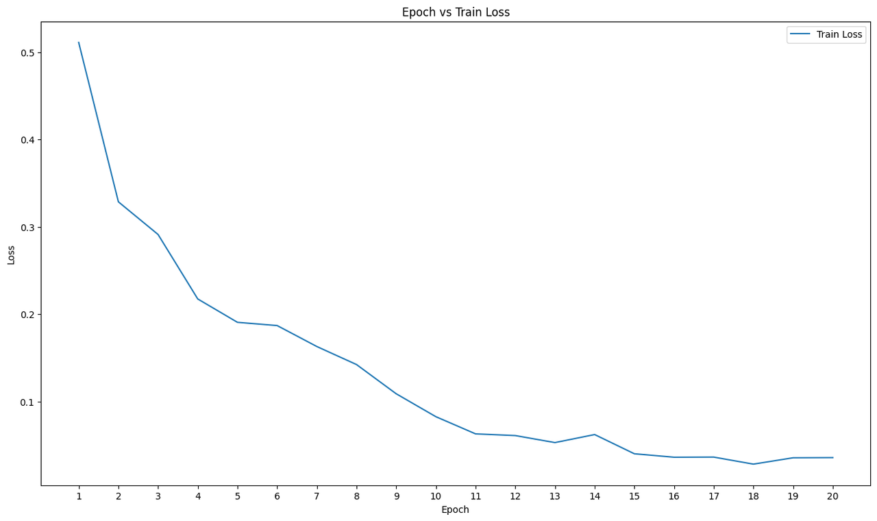
Figure 1: Training loss across epochs.
Testing Loss
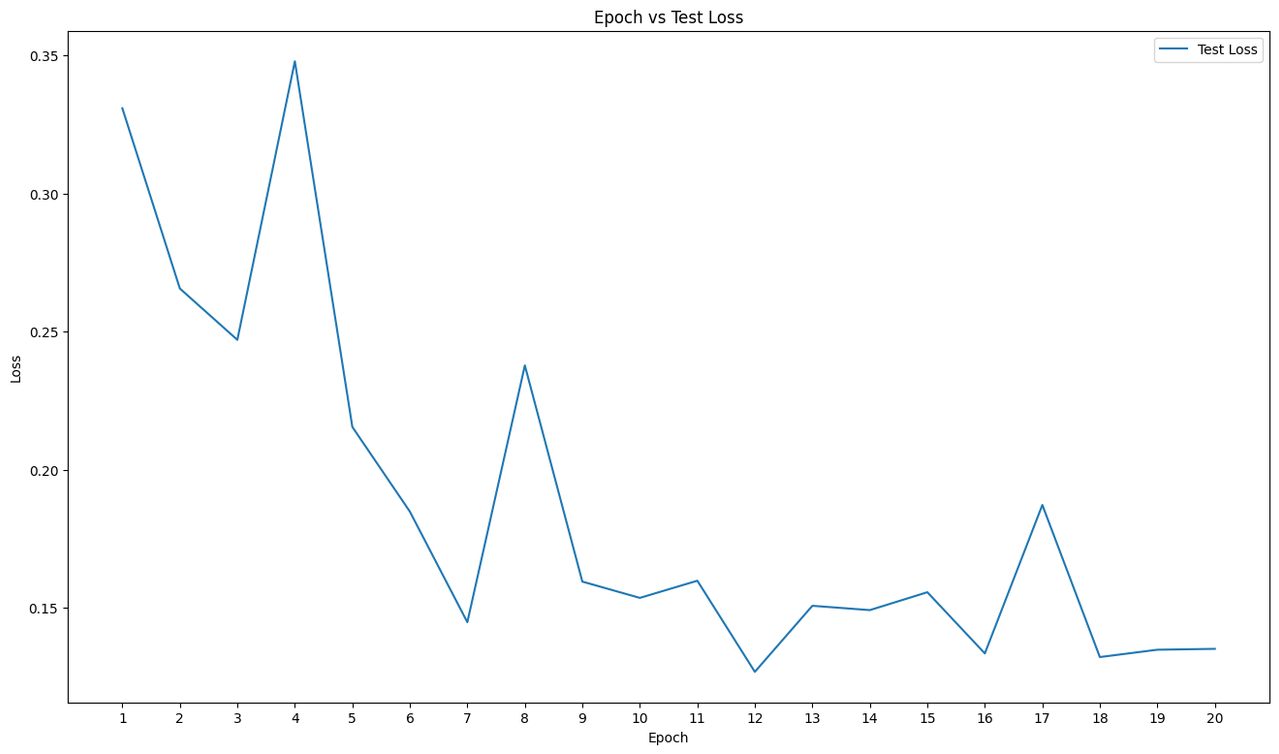
Figure 2: Testing loss across epochs.
Testing Accuracy
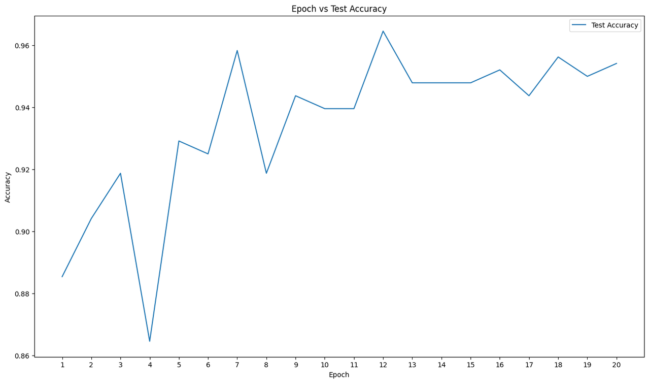
Figure 3: Test accuracy across epochs.
The combination of:
- Residual learning,
- Batch Normalization,
- Adaptive learning-rate scheduling,
- Global Average Pooling, and
- Dropout regularization,
contributed significantly to efficient optimization and strong generalization.
Classification Report
| Class | Precision | Recall | F1-Score | Support |
|---|---|---|---|---|
lung_n |
0.93 | 0.93 | 0.93 | 164 |
lung_aca |
1.00 | 1.00 | 1.00 | 167 |
lung_scc |
0.93 | 0.93 | 0.93 | 149 |
Overall Metrics
| Metric | Score |
|---|---|
| Accuracy | 0.95 |
| Macro Average | 0.95 |
| Weighted Average | 0.95 |
Confusion Matrix Analysis
Confusion Matrix
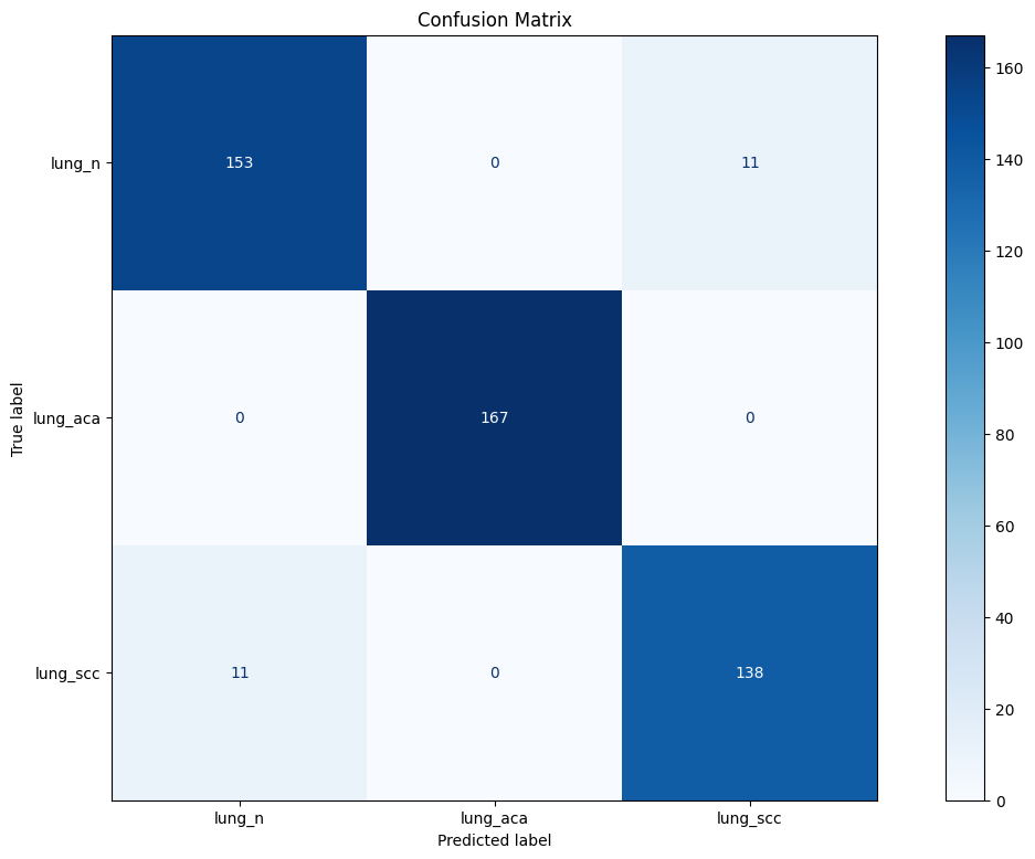
Figure 4: Confusion Matrix.
`| True Class | Correct Predictions | Total Samples | Class Accuracy |
|---|---|---|---|
lung_n |
153 | 164 | 93.29% |
lung_aca |
167 | 167 | 100.00% |
lung_scc |
138 | 149 | 92.62% |
Key Observations
Lung Adenocarcinoma (lung_aca)
The model achieved perfect classification performance for adenocarcinoma samples:
- 167 / 167 correctly classified
- No false positives
- No false negatives
- Precision = 1.00
- Recall = 1.00
- F1-score = 1.00
This demonstrates exceptionally strong feature extraction capabilities and complete separation from the remaining classes.
Lung Squamous Cell Carcinoma (lung_scc)
The model correctly classified:
138 / 149 samples
Misclassifications:
- 11 samples predicted as normal tissue.
- No confusion with adenocarcinoma samples.
This indicates strong subtype-specific feature learning and robust cancer-class separation.
Normal Lung Tissue (lung_n)
The model correctly classified:
153 / 164 samples
Misclassifications:
- 11 samples predicted as squamous cell carcinoma.
- No samples predicted as adenocarcinoma.
The absence of confusion with adenocarcinoma highlights the effectiveness of the learned feature representations.
Overall Interpretation
The confusion matrix reveals several important characteristics:
- Zero confusion involving adenocarcinoma samples.
- Misclassifications occur exclusively between normal tissue and squamous cell carcinoma.
- Both cancer classes remain distinctly separated.
Achieving perfect adenocarcinoma detection alongside more than 92% class accuracy on the remaining categories demonstrates the effectiveness of residual learning for histopathological image classification.
Dataset and Other Files
Dataset: Google Drive Dataset
Model Link Model
Colab File: Colab File
Future Work
Several directions can further enhance this work:
Data Augmentation
Future experiments will explore:
- Random rotations
- Horizontal and vertical flips
- Color jittering
- Elastic transformations
- Stain normalization methods
Increasing sample diversity may further improve robustness and reduce confusion between normal tissue and squamous cell carcinoma.
Advanced Training Strategies
Potential improvements include:
- MixUp and CutMix augmentation.
- Cross-validation experiments.
- Class-balanced sampling.
- Label smoothing techniques.
- Attention modules such as CBAM and SE blocks.
- Transfer learning from large-scale medical imaging models.
Model Interpretability
Future work may also incorporate:
- Grad-CAM visualizations.
- Feature activation analysis.
- Explainable AI (XAI) methods for clinical interpretability.
Deployment
Planned deployment directions include:
- Publishing pretrained weights on Hugging Face.
- Building an interactive inference interface.
- Packaging lightweight deployment pipelines for research and educational use.
Tech Stack
- Python
- PyTorch
- Torchvision
- NumPy
- Pandas
- Matplotlib
- Scikit-learn
Conclusion
This project demonstrates that a lightweight, custom-designed Residual CNN can effectively classify lung histopathological images with strong generalization and excellent class-wise performance.
By incorporating residual learning, batch normalization, adaptive optimization, global average pooling, and regularization techniques, the model achieved:
- 96.46% peak validation accuracy
- 0.1268 best validation loss
- 100% adenocarcinoma detection (167/167 samples)
- 95% overall classification accuracy
- 0.95 macro and weighted F1-scores
These results highlight the potential of residual architectures for medical image analysis and establish a strong foundation for future work involving larger datasets, advanced augmentation strategies, explainable AI techniques, and deployment-oriented applications.
Evaluation results
- Accuracy on Lung Histopathology Datasettest set self-reported0.960
- F1 Score on Lung Histopathology Datasettest set self-reported0.950