

image imagewidth (px) 1.53k 1.72k |
|---|
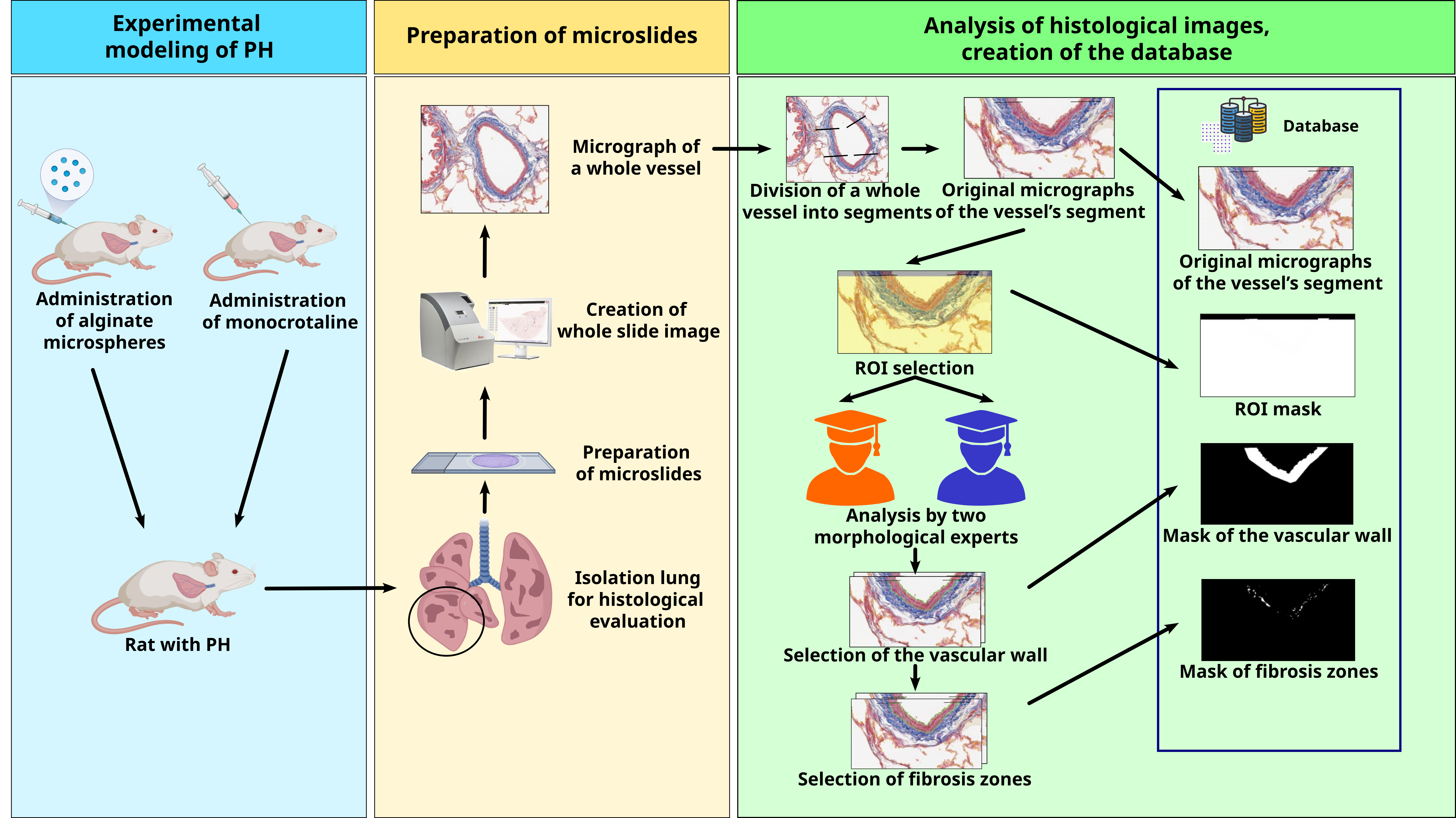
Structural remodeling of the vascular wall is a universal response to chronic damage, pressure overload or flow overload. Therefore, this process occurs in a wide range of diseases affecting both the systemic and pulmonary circulations. Vascular wall fibrosis is one of the most important factors in the pathogenesis of various types of pulmonary hypertension. In this regard quantitative assessment of collagen deposition in the vascular wall is crucial for both preclinical studies and directly affects the life prognosis for patients with pulmonary hypertension. Also, histological analysis is the gold standard for assessing vascular structural remodeling. This dataset includes 705 micrographs of vascular wall region with varying degrees of vascular wall fibrosis that developed in various types of pulmonary hypertension. All included studies were devoted to the investigation of pulmonary arterial hypertension and chronic thromboembolic pulmonary hypertension in male Wistar rats. Lung sections were stained with Picro-Mallory and digitized using a whole slide scanner. Pulmonary artery branches were subdivided into vessel segments, and exported as fixed-size micrographs. For each micrograph, the dataset provides a region-of-interest mask and two independent expert annotations, including binary masks of the vascular wall and intramural fibrosis zones. The presented database will be useful for the development of new software solutions for the analysis of histological micrographs.
The dataset comprises 705 vessel images acquired in four distinct studies. Images are named using the {PC}\GN}-{AID}-L{LL}_{FID}.png convention, where {PC} denotes the project code, {GN} is a group number, {AID} represents an animal identification, {LL} is lung section level and {FID} indicates a frame identification. For example, the file name RA1-1-L3_5.1x40.png denotes the frame ``5.1x40'', third section level of the lung, acquired from a sample of animal 1 in group 1 of project RA.
The raw color micrographs retained an 8-bit depth per RGB channel (24-bit color depth) and saved as PNG images. All masks are single channel grayscale PNG images with values 0 and 255 for background and foreground respectively.
See details:
Karpov, A.A., Sinitca, A., Isakova, N.P. et al. Microscopy Image Dataset of pulmonary vessels for Quantitative assessment of fibrosis. Sci Data (2026). https://doi.org/10.1038/s41597-026-08214-y
- Downloads last month
- 281