

CENet: Context Enhancement Network for Medical Image Segmentation
Paper • 2505.18423 • Published



Official pretrained weights for CENet (MICCAI 2025, early accept, top 9%), a 2D segmentation network for abdominal CT, cardiac MRI and dermoscopy images.
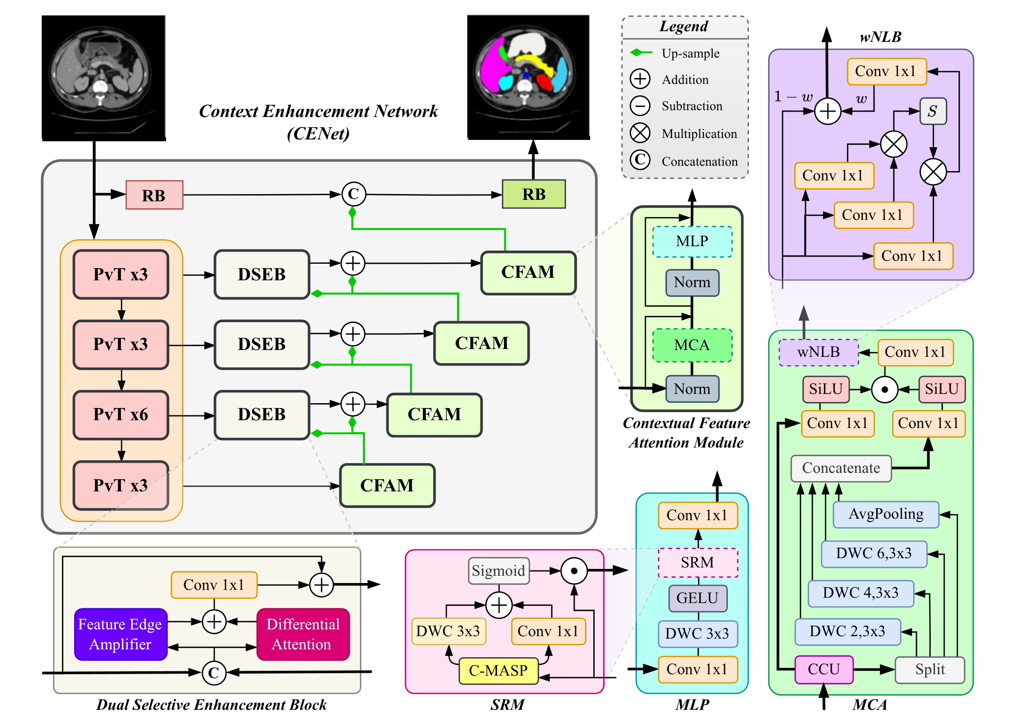
CENet uses a PVTv2-B2 encoder and adds two modules:
You do not need to clone anything. PyTorch Hub fetches the model code from GitHub, and the weights are downloaded from this repository.
pip install torch torchvision timm==1.0.16 monai==1.4.0 "numpy<2" scipy SimpleITK huggingface_hub
The predictor applies the same preprocessing as in training and returns a mask with the size of your input.
import torch

# Skin lesion (dermoscopy image)
skin = torch.hub.load("xmindflow/cenet", "predictor", dataset="ham10000", trust_repo=True)
mask = skin.predict("lesion.jpg")            # (H, W): 0 = background, 1 = lesion

# Abdominal CT (NIfTI volume, processed slice by slice)
ct = torch.hub.load("xmindflow/cenet", "predictor", dataset="synapse", trust_repo=True)
mask = ct.predict("abdomen_ct.nii.gz")       # (D, H, W)
print(ct.classes)                            # label names, index = label value
predict accepts a file path (.png, .jpg, .npy, .nii, .nii.gz), a PIL image or a NumPy array. It runs on the GPU if one is available.
model = torch.hub.load("xmindflow/cenet", "cenet", dataset="synapse", trust_repo=True)
x = torch.randn(1, 1, 224, 224)              # one preprocessed slice
with torch.no_grad():
    logits = model(x)                        # (1, 9, 224, 224)
pred = logits.argmax(dim=1)
The original model reported in the paper for Synapse is available as torch.hub.load("xmindflow/cenet", "cenet_org", trust_repo=True).
dataset |
Task | Input | Labels | File |
|---|---|---|---|---|
synapse |
Abdominal CT, 8 organs | 1 × 224 × 224 | 0 background, 1 aorta, 2 gallbladder, 3 left kidney, 4 right kidney, 5 liver, 6 pancreas, 7 spleen, 8 stomach | synapse_2418962.pth |
acdc |
Cardiac MRI | 1 × 224 × 224 | 0 background, 1 right ventricle, 2 myocardium, 3 left ventricle | acdc_2418732.pth |
ham10000 |
Skin lesion | 3 × 224 × 224 | 0 background, 1 lesion | ham10k_2418602.pth |
ph2 |
Skin lesion | 3 × 224 × 224 | 0 background, 1 lesion | ph2_2418571.pth |
cenet_org |
Abdominal CT, original paper model | 1 × 224 × 224 | same as synapse |
synapse_cenet_org_2352272.pth |
Each file is a plain PyTorch state_dict (about 130 MB).
The predictor does this for you. If you call the model directly, prepare inputs the same way:
Predictions are made at 224 × 224 and resized back with nearest-neighbor interpolation.
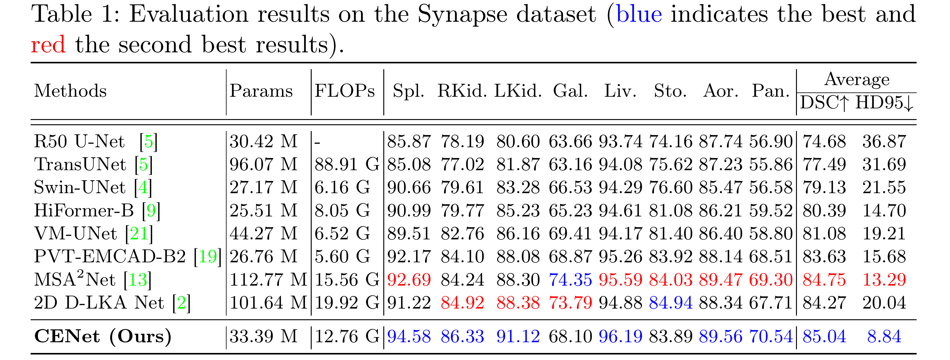
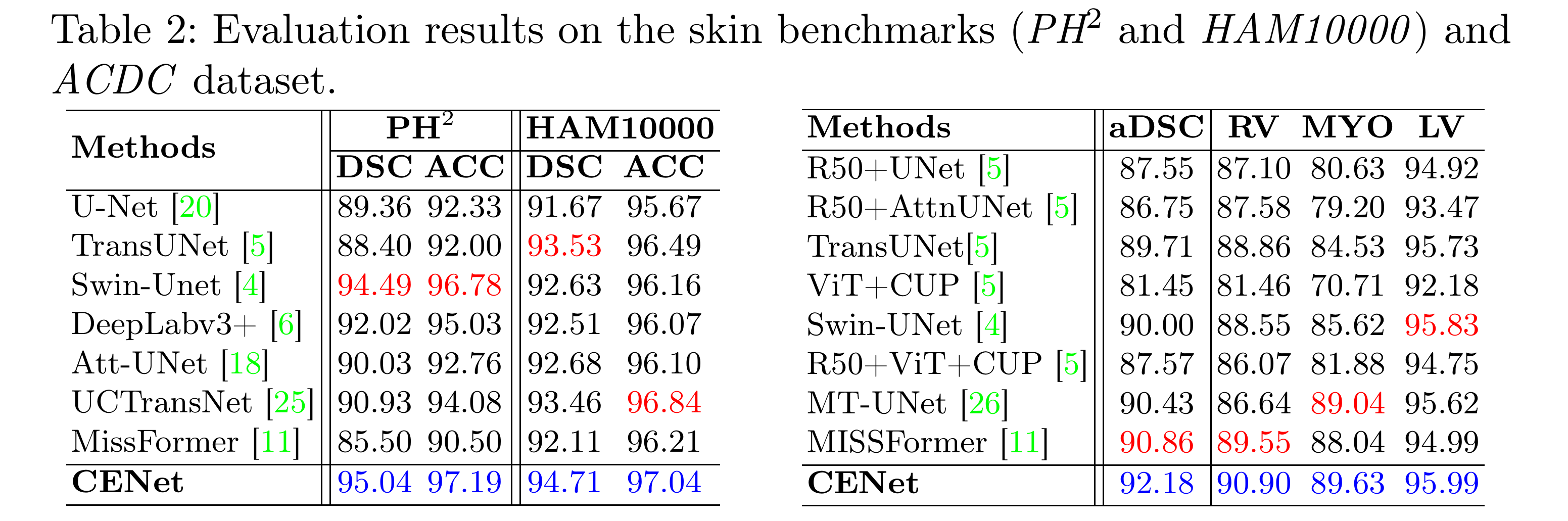
To reproduce the paper's numbers, use the evaluation scripts in the GitHub repository. The README explains how to get and prepare each dataset.
hf download afshinbigboy/CENet --include "*.pth" --local-dir weights
or in Python:
from huggingface_hub import hf_hub_download
path = hf_hub_download(repo_id="afshinbigboy/CENet", filename="synapse_2418962.pth")
These models are for research only. They are not a medical device and must not be used for diagnosis or treatment decisions. Each model was trained on a single public dataset and may perform poorly on images from other scanners, protocols or populations.
The code and weights are released under the MIT license. The training datasets have their own terms (for example, HAM10000 is CC BY-NC 4.0), so please also respect the license of each dataset.
If you use CENet, please cite:
@InProceedings{BozAfs_CENet_MICCAI2025,
  author    = {Bozorgpour, Afshin and Kolahi, Sina Ghorbani and Azad, Reza and Hacihaliloglu, Ilker and Merhof, Dorit},
  title     = {{CENet}: Context Enhancement Network for Medical Image Segmentation},
  booktitle = {Medical Image Computing and Computer Assisted Intervention -- MICCAI 2025},
  series    = {Lecture Notes in Computer Science},
  volume    = {15960},
  pages     = {120--129},
  year      = {2025},
  publisher = {Springer Nature Switzerland},
  address   = {Cham},
  doi       = {10.1007/978-3-032-04927-8_12}
}