Description
A pre-trained model for inferencing volumetric (3D) kidney substructures segmentation from contrast-enhanced CT images (Arterial/Portal Venous Phase).
A tutorial and release of model for kidney cortex, medulla and collecting system segmentation.
Authors: Yinchi Zhou (yinchi.zhou@vanderbilt.edu) | Xin Yu (xin.yu@vanderbilt.edu) | Yucheng Tang (yuchengt@nvidia.com) |
Model Overview
A pre-trained UNEST base model [1] for volumetric (3D) renal structures segmentation using dynamic contrast enhanced arterial or venous phase CT images.
Data
The training data is from the [ImageVU RenalSeg dataset] from Vanderbilt University and Vanderbilt University Medical Center. (The training data is not public available yet).
- Target: Renal Cortex | Medulla | Pelvis Collecting System
- Task: Segmentation
- Modality: CT (Artrial | Venous phase)
- Size: 96 3D volumes
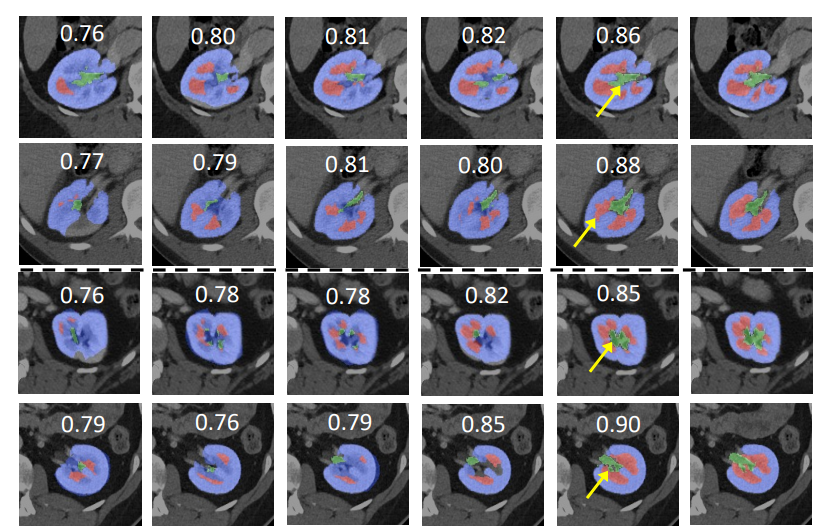
The data and segmentation demonstration is as follow:
Method and Network
The UNEST model is a 3D hierarchical transformer-based semgnetation network.
Training configuration
The training was performed with at least one 16GB-memory GPU.
Actual Model Input: 96 x 96 x 96
Input and output formats
Input: 1 channel CT image
Output: 4: 0:Background, 1:Renal Cortex, 2:Medulla, 3:Pelvicalyceal System
Performance
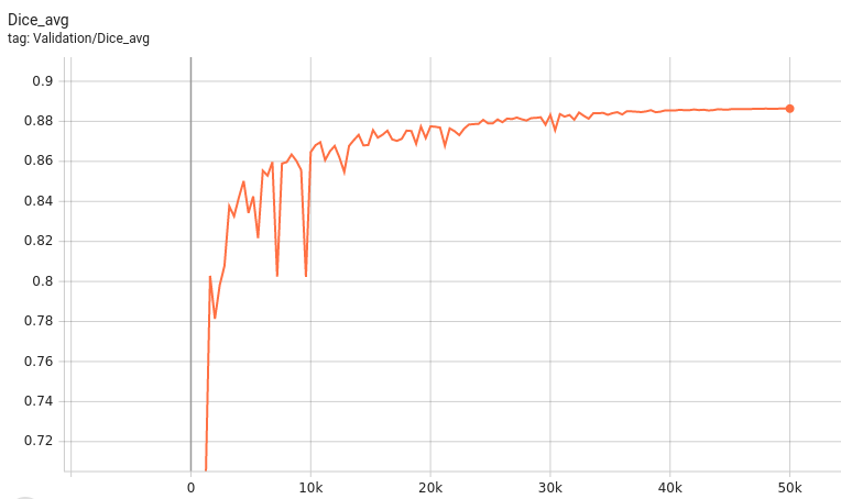
A graph showing the validation mean Dice for 5000 epochs.
This model achieves the following Dice score on the validation data (our own split from the training dataset):
Mean Valdiation Dice = 0.8523
Note that mean dice is computed in the original spacing of the input data.
commands example
Download trained checkpoint model to ./model/model.pt:
Add scripts component: To run the workflow with customized components, PYTHONPATH should be revised to include the path to the customized component:
export PYTHONPATH=$PYTHONPATH:"'<path to the bundle root dir>/scripts'"
Execute inference:
python -m monai.bundle run evaluating --meta_file configs/metadata.json --config_file configs/inference.json --logging_file configs/logging.conf
More examples output
Disclaimer
This is an example, not to be used for diagnostic purposes.
References
[1] Yu, Xin, Yinchi Zhou, Yucheng Tang et al. "Characterizing Renal Structures with 3D Block Aggregate Transformers." arXiv preprint arXiv:2203.02430 (2022). https://arxiv.org/pdf/2203.02430.pdf
[2] Zizhao Zhang et al. "Nested Hierarchical Transformer: Towards Accurate, Data-Efficient and Interpretable Visual Understanding." AAAI Conference on Artificial Intelligence (AAAI) 2022


