
datasets:
  - pubmed
language:
  - en
metrics:
  - bleu
  - exact_match
  - sacrebleu
  - rouge
tags:
  - medical
  - dialog
  - arxiv:2209.12177
widget:
  - text: >-
      The liver is normal in size and with normal parenchymal echogenicity with
      no sign of space-occupying lesion or bile ducts dilatation. GB is well
      distended with no stone or wall thickening. The spleen is normal in size
      and parenchymal echogenicity with no sign of space-occupying lesion.
      visualized parts of the pancreas and para-aortic area are unremarkable.
      Both kidneys are normal in size with normal cortical parenchymal
      echogenicity with no sign of the stone, stasis, or perinephric collection.
      ureters are not dilated. The urinary bladder is empty so evaluation of
      pelvic organs is not possible. no free fluid is seen in the abdominopelvic
      cavity.
    example_title: Sample 1
  - text: >-
      Liver is normal in size, with normal echogenicity with no sign of space
      occupying lesion. GB is semi distended with two stones up to 8mm in size
      with a rim of pericholecystic fluid and positive murphy sign. CBD is
      normal. Spleen is moderately enlarged with normal parenchymal echo with no
      S.O.L. Pancreas cannot be evaluated due to severe gas shadow. RT. and LT.
      kidneys are normal in size with increase parenchymal echogenicity with no
      sign of stone, stasis or perinephric collection with two cortical cystsin
      the upper pole of left kidney. Urinary bladder is mildly distended.
      Moderate free fluid is seen in the abdominopelvic cavity at present time.
    example_title: Sample 2
inference:
  parameters:
    repetition_penalty: 1
    num_beams: 5
    early_stopping: true
    max_length: 350
license: mit
ReportQL — Application of Deep Learning in Generating Structured Radiology Reports: A Transformer-Based Technique
Seyed Ali Reza Moezzi, Abdolrahman Ghaedi, Mojdeh Rahmanian, Seyedeh Zahra Mousavi, Ashkan Sami
[paper] [arXiv] [dataset] [project page]
Introduction
This repository is code release for Application of Deep Learning in Generating Structured Radiology Reports: A Transformer-Based Technique
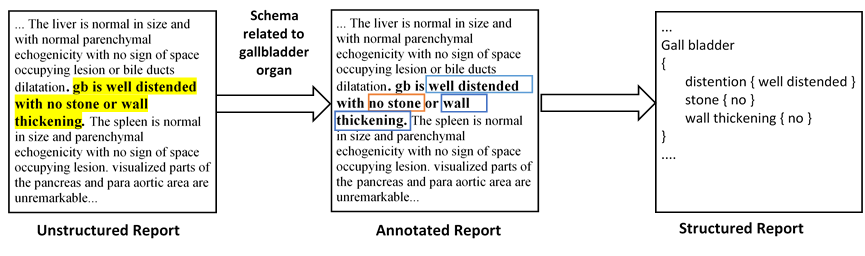
Since radiology reports needed for clinical practice and research are written and stored in free-text narrations, extraction of relative information for further analysis is difficult. In these circumstances, natural language processing (NLP) techniques can facilitate automatic information extraction and transformation of free-text formats to structured data. In recent years, deep learning (DL)-based models have been adapted for NLP experiments with promising results. Despite the significant potential of DL models based on artificial neural networks (ANN) and convolutional neural networks (CNN), the models face some limitations to implement in clinical practice. Transformers, another new DL architecture, have been increasingly applied to improve the process. Therefore, in this study, we propose a transformer-based fine-grained named entity recognition (NER) architecture for clinical information extraction. We collected 88 abdominopelvic sonography reports in free-text formats and annotated them based on our developed information schema. The text-to-text transfer transformer model (T5) and Scifive, a pre-trained domain-specific adaptation of the T5 model, were applied for fine-tuning to extract entities and relations and transform the input into a structured format. Our transformer-based model in this study outperformed previously applied approaches such as ANN and CNN models based on ROUGE-1, ROUGE-2, ROUGE-L, and BLEU scores of 0.816, 0.668, 0.528, and 0.743, respectively, while providing an interpretable structured report.
Dataset
Our annotated dataset used in the paper is hosted in this repository and in Kaggle Datasets.
The data is structured as follows:
data/
├── trialReport
│   └── ReportQL
│       ├── Schemas
│       │   └── organs
│       │       └── simpleSchema.json
│       └── dataset
│           ├── test.csv
│           ├── train_orig.csv
│           └── training.csv
The train_orig.csv is our original training set. You can find our synthetic dataset and test set in training.csv and test.csv file.
Information schema used for annotating reports can be found in simpleSchema.json
Setup
Setting up for this project involves installing dependencies.
Setting up environments and Installing dependencies
virtualenv .venv
source .venv/bin/activate
Installing dependencies
To install all the dependencies, please run the following:
pip install -r requirements.txt
Fine-tuning
To start fine-tuning language model, run:
python script/fit.py
Testing
For getting test results on our test set, run:
python script/test.py
Inference
We prepared a jupyter notebook for Inference.
Fine-tuned Model
Our fine-tuned ReportQL weights can be accessed on 🤗 HuggingFace.
- ReportQL: base
License
Please see the LICENSE file for details.
Citation
If you find our work useful in your research, please consider citing us:
@article{moezzi2022application,
  title={Application of Deep Learning in Generating Structured Radiology Reports: A Transformer-Based Technique},
  author={Moezzi, Seyed Ali Reza and Ghaedi, Abdolrahman and Rahmanian, Mojdeh and Mousavi, Seyedeh Zahra and Sami, Ashkan},
  journal={Journal of Digital Imaging},
  pages={1--11},
  year={2022},
  publisher={Springer}
}